
ADVANCED OCULAR SURFACE & DRY EYE ASSESSMENT
Precision Diagnosis
Inflammatory Profiling
Targeted Treatment

Understanding the Root Cause of Ocular Surface Disease
Dry eye disease and ocular surface disorders are among the most common causes of ocular discomfort, fluctuating vision, contact lens intolerance, and reduced quality of life. However, dry eye is not a single disease. It is a complex condition involving structural changes, inflammation, immune dysregulation, and tear film instability.
Our Advanced Ocular Surface & Dry Eye Assessment combines state-of-the-art imaging, tear biomarker analysis, and targeted therapies to identify the underlying mechanisms driving each patient's symptoms and to develop a personalized treatment strategy.
COMPREHENSIVE DIAGNOSTIC ASSESSMENT
01
Structural Analysis
Meibography
Meibography is a non-invasive imaging technique that allows direct visualization of the meibomian glands, which produce the lipid layer of the tear film and play a critical role in maintaining ocular surface health.
Clinical Benefits
- Early detection of Meibomian Gland Dysfunction (MGD)
- Identification of gland dropout and atrophy
- Objective assessment of gland structure
- Detection of changes before irreversible damage occurs
- Monitoring of disease progression and treatment response
- Identification of patients who may benefit from IPL therapy and gland expression
Why It Matters
Meibomian Gland Dysfunction is the leading cause of evaporative dry eye disease. Structural gland loss can occur long before symptoms become severe. Early identification enables earlier intervention and improved long-term outcomes.
03
INFLAMMATORY PROFILING
Advanced Tear Immunoanalysis
Tear fluid contains valuable biological information about the inflammatory and immune status of the ocular surface. Our biomarker panel provides objective insight into disease activity and helps guide treatment decisions.
02
COMPREHENSIVE CLINICAL EVALUATION
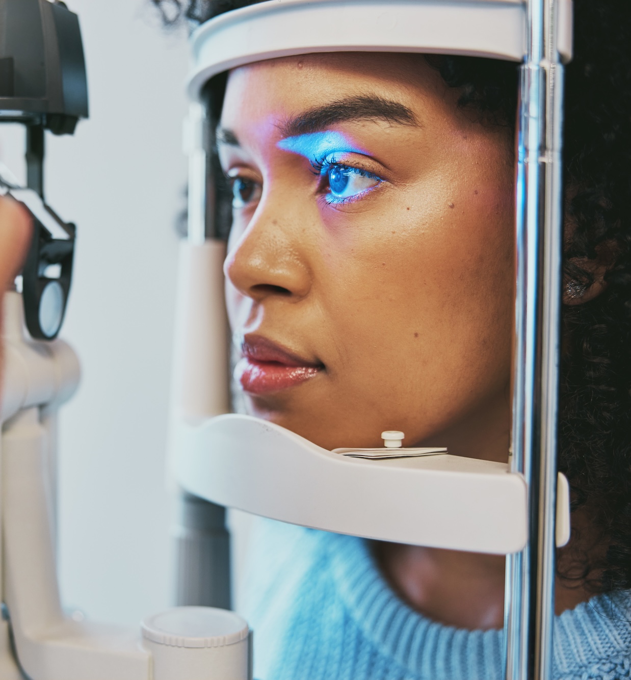
Slit Lamp Examination
A detailed slit lamp assessment provides a thorough evaluation of the eyelids, tear film, and ocular surface.
The examination includes assessment of:
- Eyelid margin health
- Blepharitis
- Meibomian gland function
- Tear film quality and stability
- Corneal and conjunctival integrity
- Ocular surface staining
- Telangiectatic vessels
- Signs of chronic inflammation
Benefits
- Correlation of symptoms with clinical findings
- Disease severity grading
- Identification of contributing factors
- Development of a personalized management plan

MMP-9 (Matrix Metalloproteinase-9)
MMP-9 is one of the most extensively validated biomarkers of ocular surface inflammation.
Elevated MMP-9 May Indicate
- Active ocular surface inflammation
- Moderate to severe dry eye disease
- Corneal epithelial stress and damage
- Persistent inflammatory activity
Clinical Benefits
- Objective confirmation of inflammation
- Identification of patients who may benefit from anti-inflammatory therapies
- Monitoring of treatment effectiveness
- More precise therapeutic decision-making

Tear IgE
Immunoglobulin E (IgE) isan important biomarker associated with allergic inflammation.
Elevated Tear IgE May Suggest
- Allergic conjunctivitis
- Ocular hypersensitivity
- Mixed allergic and dry eye disease
- Subclinical allergic inflammation
Clinical Benefits
- Differentiation between allergic and non-allergic inflammation
- Improved treatment selection
- Reduced reliance on empirical therapy
- Better long-term symptom control
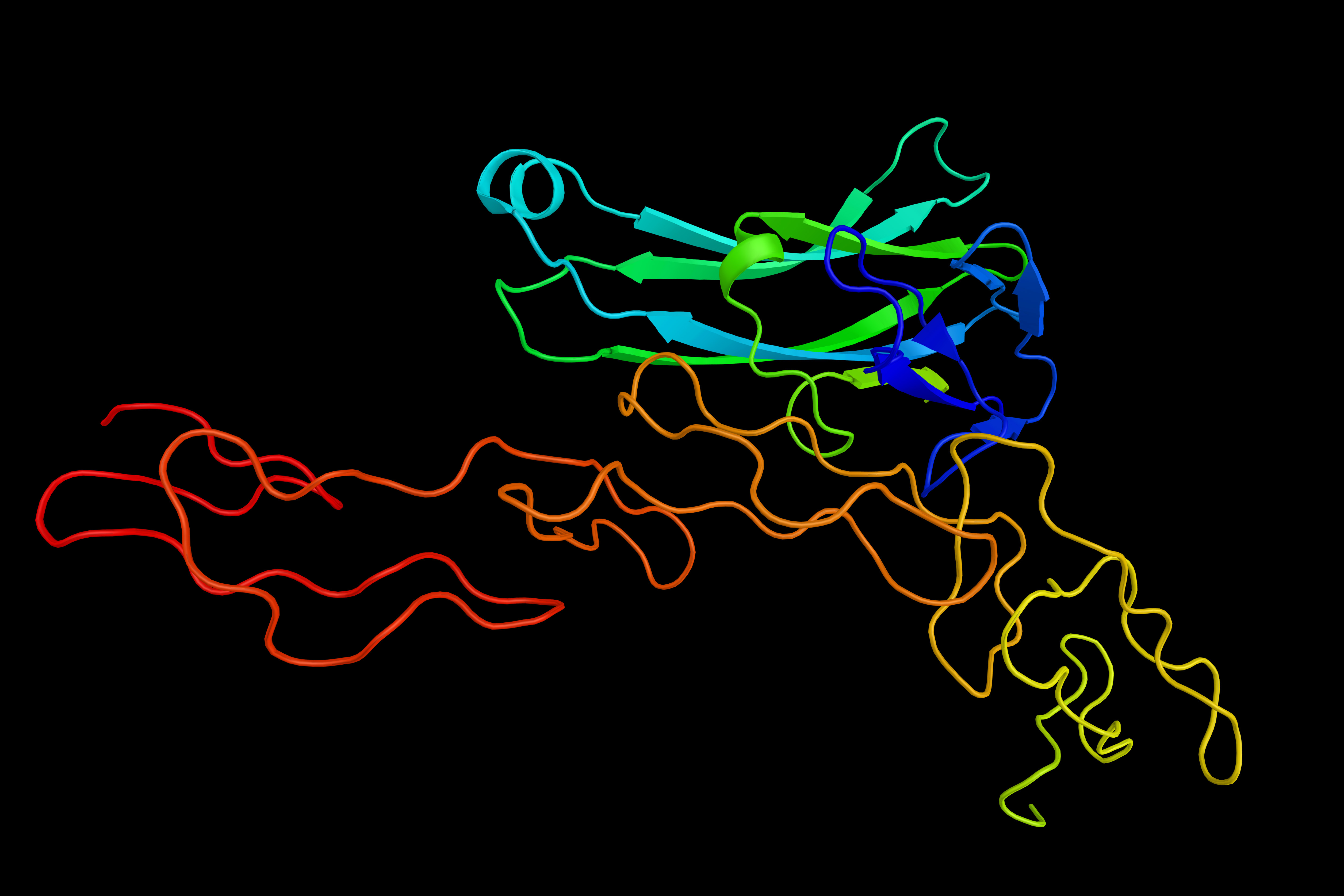
LYMPHOTOXIN-ALPHA (LTα)
A Novel Biomarker of Ocular Surface Immune Activity
Lymphotoxin-Alpha (LTα), also known as TNF-β, is a cytokine involved in immune regulation and chronic inflammatory responses. Emerging evidence suggests that LTα may play an important role in immune-mediated ocular surface disease.
Unlike conventional dry eye testing, LTα provides insight into the immune environment of the ocular surface and may help identify patients with a more inflammatory disease phenotype.
Why LTα Matters
Dry eye disease is increasingly recognized as an inflammatory disorder rather than simply a deficiency of tears.
While traditional assessments focus on tear quantity and ocular surface damage, LTα provides additional information regarding the immune activity occurring on the ocular surface.
Emerging evidence suggests that elevated LTα levels may be associated with enhanced lymphocyte-mediated inflammation and chronic immune activation. This information may help identify patients whose symptoms are driven by inflammatory mechanisms that are not readily apparent through routine clinical examination alone.
Although LTα is not a diagnostic marker for autoimmune disease, it may provide valuable information regarding the inflammatory phenotype of ocular surface disease and may support decisions regarding further investigation, treatment selection, and long-term monitoring.
Clinical Relevance of Elevated LTα
Elevated LTα levels may indicate:
- Increased immune activation within the ocular surface environment
- Chronic inflammatory activity
- Lymphocyte-mediated inflammation
- Greater risk of persistent symptoms
- A more treatment-resistant inflammatory phenotype
In some patients, elevated LTα levels may coexist with systemic immune-mediated disorders such as:
- Sjögren's Syndrome
- Rheumatoid Arthritis
- Systemic Lupus Erythematosus
- Autoimmune Thyroid Disease
- Other connective tissue disorders
- LTα should always be interpreted within the context of the patient's clinical findings and medical history and should not be considered a standalone diagnostic marker of autoimmune disease.
TARGETED TREATMENT OPTIONS
01
Meibomian Gland Expression (54)
Meibomian gland expression removes retained gland contents and helps restore normal gland function.
Benefits
- Clears obstructed glands
- Improves meibum quality
- Restores lipid layer function
- Reduces tear evaporation
- Improves tear film stability
- Enhances ocular comfort
Expected Outcomes
- Reduced dryness
- Reduced irritation
- Improved visual stability
- Better contact lens tolerance
- Greater day-to-day comfort
02
INTENSE PULSED LIGHT (IPL) THERAPY
(£1000 package or £300 per session)
Advanced Treatment for Meibomian Gland Dysfunction and Ocular Surface Inflammation
Intense Pulsed Light (IPL) therapy has become one of the most effective treatments available for chronic dry eye disease associated with Meibomian Gland Dysfunction.
Beyond improving meibomian gland function, IPL is increasingly recognized as a disease-modifying therapy capable of reducing chronic ocular surface inflammation and improving tear film homeostasis.
How IPL light therapy works
Reduces Inflammation
IPL decreases inflammatorymediators within the eyelids and ocular surface, helping to break the cycle ofchronic inflammation.
Eliminates Abnormal Blood Vessels
Telangiectatic vesselscontribute to ongoing inflammation through the release of inflammatorymediators. IPL selectively targets these vessels, reducing their inflammatoryinfluence.
Improves Meibomian Gland Function
The thermal energygenerated by IPL helps liquefy thickened meibum and improves gland secretion.
Reduces Bacterial Load
IPL decreases bacterialcolonization associated with chronic blepharitis and eyelid inflammation.
Helps Control Demodex
IPL may reduce Demodexpopulations, a common contributor to chronic eyelid inflammation and ocularirritation.
Who Can Benefit from IPL Therapy?
- Dry Eye Disease
- Meibomian Gland Dysfunction (MGD)
- Ocular Rosacea
- Blepharitis
- Recurrent Styes (Chalazia)
- Demodex Eyelid Infestation
- Red, Irritated Eyes
- Contact Lens Discomfort
- Fluctuating or Blurred Vision Related to Dry Eye
- Chronic Eyelid Inflammation
- Patients Requiring Frequent Artificial Tear Use
- Persistent Symptoms Despite Conventional Dry Eye Treatments
- Chronic Ocular Surface Inflammation
IPL is particularlyeffective for patients with Meibomian Gland Dysfunction and Ocular Rosacea, twoof the most common causes of chronic dry eye symptoms.
03
Scleral Lenses for SevereDry Eye Disease
For patients with moderate to severe dry eye disease, scleral lenses can provide both visual rehabilitation and therapeutic protection of the ocular surface.
Unlike conventional contact lenses, scleral lenses vault over the cornea and create a fluid-filled reservoir that continuously bathes the ocular surface throughout the day. This protective layer helps reduce symptoms, improve comfort, and support ocular surface healing.
Benefits of Scleral Lenses
- Continuous hydration of the ocular surface
- Reduced dryness, burning, and irritation
- Protection of the cornea from environmental exposure
- Improved visual quality and stability
- Enhanced comfort in severe ocular surface disease
- Support of corneal healing and recovery
Particularly Beneficial For
- Severe Dry Eye Disease
- Sjögren's Syndrome
- Ocular Surface Disease
- Neurotrophic Keratopathy
- Exposure Keratopathy
- Patients with persistent symptoms despite conventional therapies
For selected patients, scleral lenses can be a highly effective adjunctive treatment alongside IPL, meibomian gland therapy, and targeted anti-inflammatory management.

SPECIALIST SCLERAL LENS SERVICE
£98 – fitting, £610 per lens
Vision Rehabilitation and Ocular Surface Protection, Restoring Vision, Comfort, and Quality of Life
Scleral contact lenses represent one of the most significant advances in modern specialty eye care. Unlike conventional contact lenses, scleral lenses vault over the cornea and rest on the sclera (the white part of the eye), creating a fluid-filled reservoir that provides exceptional vision, comfort, and protection for the ocular surface.
They are often the treatment of choice for patients with irregular corneas, severe dry eye disease, and complex ocular surface disorders where glasses or conventional contact lenses fail to provide satisfactory results.
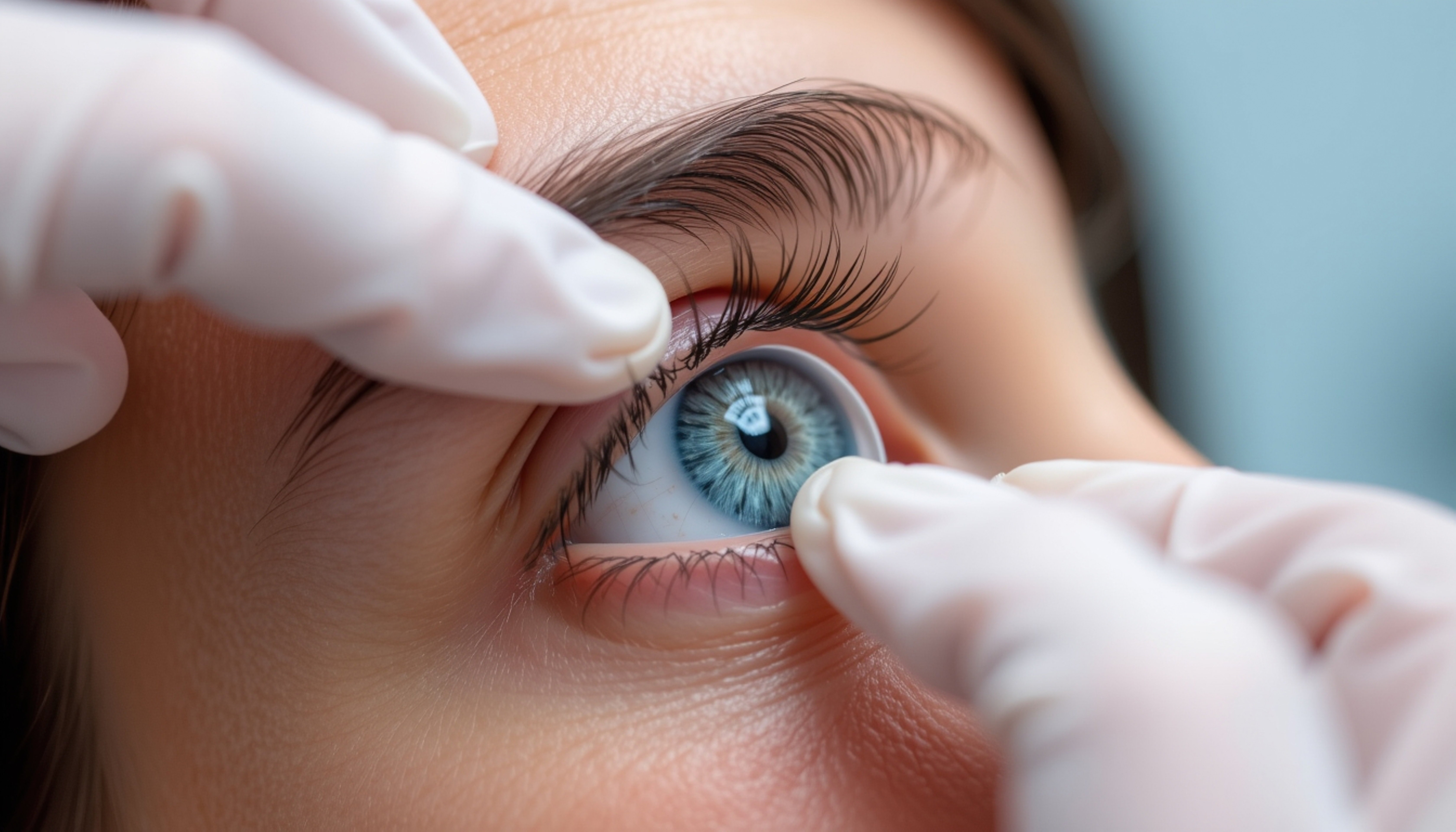
WHAT ARE SCLERAL LENSES?
Scleral lenses are large-diameter custom-designed gas permeable lenses that:
- Vault completely over the cornea
- Create a protective fluid reservoir over the ocular surface
- Provide a smooth optical surface
- Protect the cornea from mechanical irritation
- Deliver exceptional visual quality and comfort
Because each lens is individually designed, scleral lenses can be tailored to the unique shape and needs of every eye.
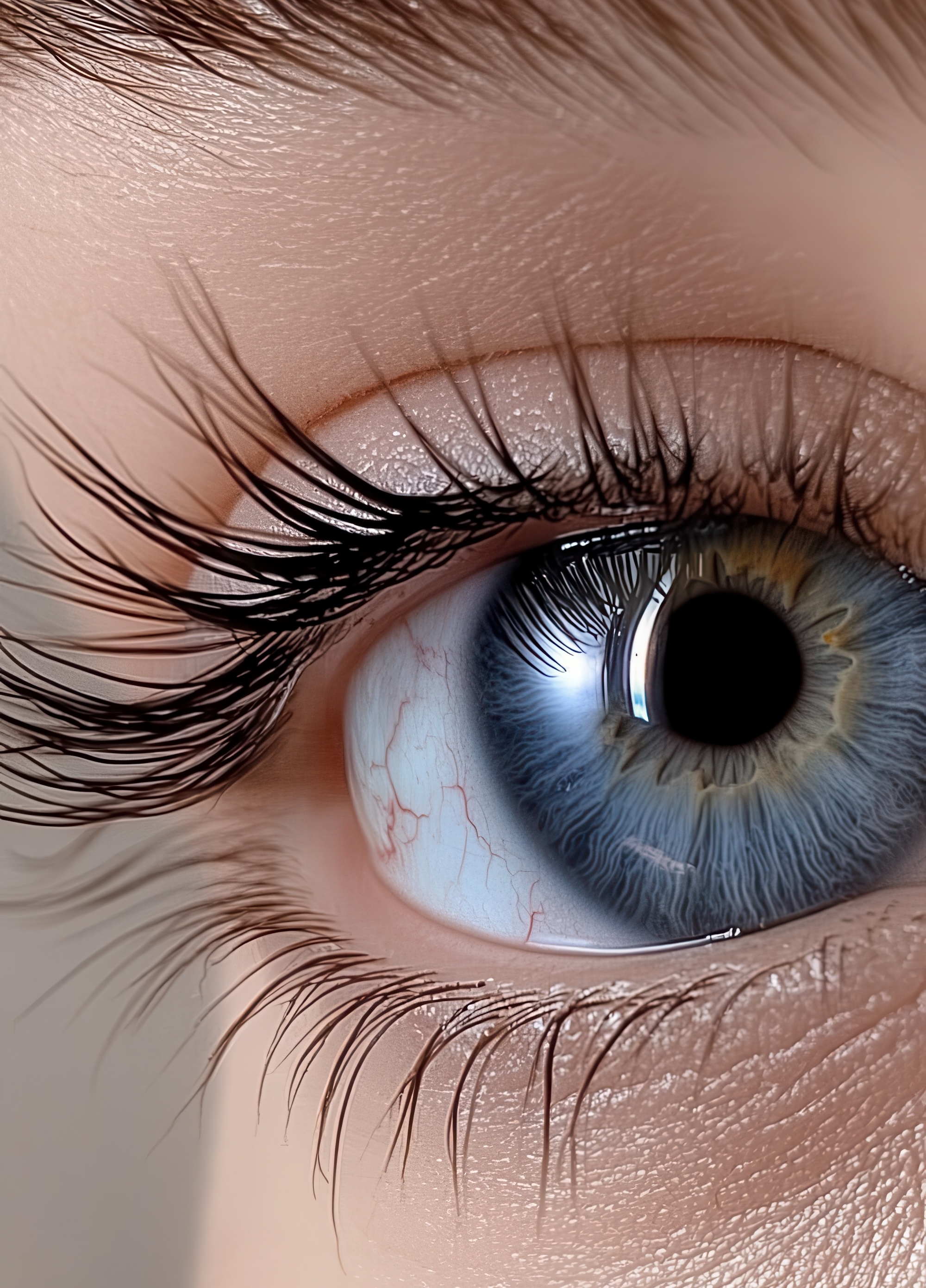
BENEFITS OF SCLERAL LENSES
Exceptional Vision
By masking corneal irregularities, scleral lenses create a perfectly smooth optical surface, often providing vision that cannot be achieved with glasses or standard contact lenses.
Patients frequently experience:
- Sharper vision
- Reduced glare and halos
- Improved contrast sensitivity
- More stable vision throughout the day
- Better night-time vision
Superior Comfort
Unlike traditional rigid contact lenses, scleral lenses do not touch the cornea.
The fluid reservoir cushions and protects the eye, making scleral lenses remarkably comfortable even for patients who have previously been unable to tolerate contact lenses.
Ocular Surface Protection
The fluid layer beneath the lens continuously bathes the ocular surface throughout the day.
Benefits include:
- Improved hydration
- Reduced friction from blinking
- Protection of fragile corneal tissue
- Enhanced ocular comfort
- Reduced symptoms of dryness and irritation
WHO CAN BENEFIT FROM SCLERAL LENSES?
Keratoconus
Scleral lenses are widely regarded as one of the most effective non-surgical options for visual rehabilitation in keratoconus.
Pellucid Marginal Degeneration
Provides excellent visual correction for highly irregular corneas.
Post-Corneal Transplant Patients
Improves vision when glasses or standard contact lenses are insufficient.
Post-Refractive Surgery Corneal Irregularity
Suitable for patients experiencing visual distortion after LASIK, PRK, RK, or other refractive procedures.
Corneal Scarring
Can significantly improve visual quality by masking irregular corneal surfaces.
Severe Dry Eye Disease
Provides continuous hydration and protection to the ocular surface.
Sjögren's Syndrome
Offers substantial symptom relief and ocular surface protection in severe dry eye associated with autoimmune disease.
Ocular Surface Disease
Useful in a range of inflammatory and cicatrizing disorders affecting the cornea and conjunctiva.
Exposure Keratopathy
Protects the cornea in patients with incomplete eyelid closure.
Neurotrophic Keratopathy
Supports corneal healing and protects vulnerable corneal tissue.
Stevens-Johnson Syndrome can provide both visual rehabilitation and significant symptom relief.
Graft-versus-Host Disease (GVHD)
Improves comfort and protects the ocular surface in severe ocular involvement.

Our fitting process
Successful scleral lenswear depends on precise customization and expert fitting.
Our comprehensive assessment includes:
- Detailed ocular history
- Corneal topography and tomography
- Evaluation of ocular surface health
- Tear film assessment
- Lens design selection
- Trial lens fitting
- Optical optimization
Book Your Dry Eye Assessment Today
Don’t let dry eyes affect your comfort and quality of life. Our experienced team at Matheson Optometrists & Hearing Care is here to provide personalised care and treatments to help you feel your best.
Contact us today to book your appointment at our Dry Eye Clinic and take the first step towards clearer, more comfortable vision.